The right test for the right clinical question.
No single test identifies every injury. GMS evaluates the injury history, symptoms, care already received, and existing findings to determine which diagnostic studies are clinically appropriate.
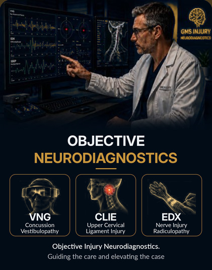
GMS performs VNG, EDX, and CLIE evaluations and coordinates imaging (MRI, CT, X-ray) and other necessary testing when indicated.
Tests GMS performs
Vestibular and neurological evaluation
VNG objectively evaluates eye movements and vestibular function. Following head or whiplash trauma, it may document central or peripheral vestibular abnormalities associated with concussion/mTBI, traumatic vestibulopathy, dizziness, vertigo, and balance dysfunction.
The findings can help define the extent of vestibular involvement and guide rehabilitation, specialist referral, treatment recommendations, and follow-up evaluation.
Upper cervical ligament injury evaluation
CLIE evaluates clinical and functional findings associated with injury to the upper cervical stabilizing ligaments following whiplash or other trauma.
The findings help determine whether additional imaging, cervical stabilization, treatment precautions, follow-up evaluation, or specialist consultation may be appropriate.
Nerve and muscle evaluation
EDX, including nerve conduction studies and EMG, objectively evaluates nerve and muscle function. It can identify and localize cervical or lumbar radiculopathy, peripheral nerve injury, entrapment neuropathy, and other neuromuscular abnormalities.
The results help establish the location and clinical significance of neurological involvement and guide treatment, injections, specialist referral, and follow-up care. When performed early enough, it may be able to establish or rule out pre-existing injury.
Imaging GMS coordinates
Detailed soft-tissue imaging
MRI provides detailed imaging of the brain, spinal cord, discs, ligaments, nerves, and other soft tissues. It may identify structural injuries that cannot be adequately evaluated by standard X-rays or CT.
MRI is important when neurological deficits, persistent symptoms, or suspected soft-tissue injuries warrant further investigation. A normal brain MRI, however, does not rule out concussion.
Alignment, motion, and bony injury
X-rays evaluate fractures, spinal alignment, joint spacing, and degenerative changes. When clinically appropriate, weight-bearing, open-mouth, and flexion-extension cervical views may provide additional information concerning alignment or abnormal motion following trauma.
These findings may help determine whether additional imaging, treatment restrictions, stabilization, or specialist referral is needed.
Acute trauma and bony detail
CT provides rapid, detailed imaging and is particularly valuable for detecting acute fractures and intracranial bleeding following trauma.
It is commonly used when urgent structural injury must be ruled out, although it is less sensitive than MRI for many ligament, disc, spinal cord, and other soft-tissue injuries. CT is not used by itself to diagnose concussion.
Why objective diagnostics matter
Objective findings help:
- Identify injuries that may otherwise be overlooked
- Support clinical causation
- Guide appropriate treatment and specialist care
- Document the extent of neurological or structural involvement
- Establish a clearer and more complete medical record
Better diagnostic information leads to better-informed care — and a stronger clinical foundation for the case.